
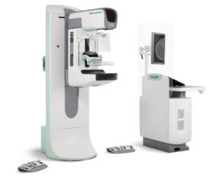
2D Mammography and 3D Mammography
The doctors and staff at Greenville Women’s Care are experienced in detecting early signs of breast cancer through 2D Mammography and 3D Mammography. Mammography is a type of imaging that uses a low-dose X-ray system to examine breasts.
A mammography exam, or mammogram, is used as a screening tool to aid in the detection and diagnosis of breast cancer in women experiencing no symptoms. They are also used to detect and diagnose breast disease in women experiencing symptoms such as a lump, pain or nipple discharge. The American College of Radiology and the Society of Breast Imaging both recommend mammography as a means of saving lives.
Mammography plays a central part in the early detection of breast cancers because it can show changes in the breast up to two years before a patient or physician can feel them. Current guidelines from the American Cancer Society recommends women begin screening mammography at age 45, and the American College of Radiology recommends screening mammography beginning at age 40, and that women have a risk assessment at age 30.
Research has shown that annual mammograms lead to early detection of breast cancers, when they are most curable and breast-conservation therapies are available.
What is the difference between 2D Mammography and 3D Mammography?
In 2D mammography, two X-ray images are taken, one from the top, and a second from the side, creating a two-dimensional image of the breast from two X-ray images of each breast.
In 3D tomosynthesis, also called digital breast tomosynthesis, digital tomosynthesis, or just tomosynthesis, more images are taken, resulting images of thin slices of the breast. Several low-dose images from different angles around the breast are used to create the 3D picture. This means that breast tissue from one side of the breast does not get in the way of the images of the other side of the breast.
3D mammography is approved by the U.S. Food and Drug Administration (FDA) and is a standard of care for breast cancer screening. A number of studies have found that 3D mammograms find more cancers than traditional 2D mammograms and also reduce the number of false positives, meaning fewer call-back for additional imaging.
A false positive is when a mammogram shows an abnormal area that looks like a cancer but turns out to be normal. Ultimately, the news is good: no breast cancer. But the suspicious area usually requires follow-up with more than one doctor, extra tests, and extra procedures, sometimes including a possible biopsy. There are psychological, physical, and economic costs that come with a false positive.
How Is the Procedure Performed?
During both 2D and 3D mammography, a specially qualified radiologic technologist will position your breast in the mammography unit. Your breast will be placed on a special platform and compressed with a paddle. The technologist will gradually compress your breast.
Breast compression is necessary in order to:
- Even out the breast thickness so that all of the tissue can be visualized.
- Spread out the tissue so that small abnormalities won’t be obscured by overlying breast tissue.
- Allow the use of a lower X-ray dose since a thinner amount of breast tissue is being imaged.
- Hold the breast still in order to eliminate blurring of the image caused by motion.
- Reduce X-ray scatter to increase sharpness of picture.
- You must hold very still and may be asked to keep from breathing for a few seconds while the X-ray picture is taken to reduce the possibility of a blurred image. The technologist will walk behind a wall or into the next room to activate the X-ray machine.
- You will be asked to change positions slightly between images. The routine views are a top-to-bottom view and an oblique side view. The process will be repeated for the other breast.
When the examination is complete, you will be asked to wait until the technologist determines that the images are of high enough quality for the radiologist to read. The examination process should take about 15 minutes.
From a patient perspective, the experience of having 2D and 3D mammograms are almost identical. In both, compression is used to immobilize the breast and to help capture images of the whole breast. 3D tomosynthesis does not require more breast compressions.
Patients should consult their physician regarding screening guidelines that are appropriate for you.